Chin J Dent Res 2026;29(3):201–215;
doi:10.3290/j.cjdr.b7093210
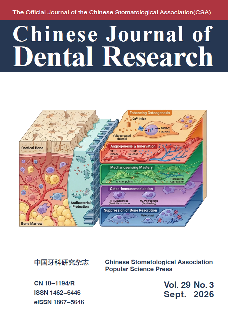
Remodelling the Electrical Microenvironment for Facilitating Bone Regeneration: a Decade of Progress
Boon Chin HENG, Yang LIU, Yun Yang BAI, Xiao Na ZHENG, Jia SONG, Qun CUI, Wan Li SONG, Xue Hui ZHANG, Xu Liang DENG
The increasing global incidence of bone injuries and degenerative diseases, often compounded by conditions like diabetes and osteoporosis, presents a significant health care challenge. Criticalsized bone defects frequently fail to heal due to compromised bioelectric microenvironments. This review summarises the conceptual framework of electric microenvironment remodelling in bone tissue, covering its physiological basis, disruption in disease and therapeutic modulation using electroactive scaffolds. The authors outline how the electric microenvironment of bone tissue is generated and continuously remodelled physiologically, analyse how its deficiency compromises regeneration and highlight strategies for dynamic regulation under pathological conditions. Restoring the bioelectric environment via electroactive scaffolds promotes bone healing by enhancing osteogenesis, angiogenesis and immunomodulation, while suppressing bone resorption. Diverse electroactive materials, including piezoelectric polymers and selfpowered nanogenerators, are critically examined. Future bone tissue engineering will likely involve smart composite electroactive scaffolds that respond to external stimuli, enabling noninvasive, dynamic and precise modulation of electrical signalling cues for effective treatment of complex bone defects.
Chin J Dent Res 2026;29(3):219–226;
doi:10.3290/j.cjdr.b7093213
Expert Consensus on the Emergency Treatment of Oral Bleeding
Chinese Stomatological Association Group Standards T/CHSA 098-2025, Society of Oral Emergency, Chinese Stomatological Association;Long JIANG, Tian Tian WU, Jun Jun ZHAO, Zhuo Jun ZHOU, Jin Heng LI, Ya Qin ZHU
Oral bleeding refers to haemorrhage occurring in the oral and maxillofacial region that does not stop spontaneously, resulting from either local factors, such as inflammation, trauma or tumours, or systemic conditions, such as haematological diseases, severe liver dysfunction or drug-related coagulopathy. Due to the abundant vascular supply in this region, blood loss can be substantial. Moreover, the admixture of blood with saliva often leads patients to mistakenly perceive the haemorrhage as more severe than it is, inducing significant anxiety. Patients with major maxillofacial trauma or systemic coagulation disorders may develop life-threatening complications including airway obstruction and hypovolemic shock. This consensus provides a framework for the emergency management of oral bleeding, encompassing pre-examination triage, principles and protocols for emergency treatment and site-specific recommendations for managing common oral haemorrhages. The aim is to standardise emergency care, assist clinicians in rapidly identifying critical conditions, achieve prompt haemostasis and ultimately improve the safety and efficiency of emergency management for patients with oral bleeding. This document is developed based on a review of the relevant literature and current clinical practices in select medical institutions and may be adapted according to the resources and circumstances of individual health care settings.
Chin J Dent Res 2026;29(3):237–246;
doi:10.3290/j.cjdr.b7093216
Effects of Central Retainer Depth on Stress Distribution and Fracture Resistance in Endocrown-restored Maxillary Premolars: 3D Finite Element and in vitro Analysis
You Sheng JIANG, Lin FENG, Jun Xin ZHU, Xiao Yan WANG, Xue Jun GAO
Objective: To explore how central retainer depth affects stress distribution and fracture resistance in endocrown-restored premolars. Methods: A 3D finite element (FE) model of an intact maxillary premolar was established as a negative control. Five endocrown-restored FE models were established, differing in the extension of the central retainer inside the pulp chamber: 0, 2, 3, 4 and 5 mm. The remaining pulp cavity was filled with composite resin. Stress distributions were analysed under oblique loads. Thirty freshly extracted human maxillary premolars were randomly divided into three groups after endodontic treatment. The extension of the central retainer was set to 0 (G0), 2 (G2) or 4 mm (G4). After endocrown manufacturing and cementation, all specimens were subjected to oblique loads in a universal testing machine until fracture occurred. Fracture resistance was analysed by one-way analysis of variance (ANOVA). Fracture patterns were analysed using a Fisher exact test (α = 0.05). Results: The FE analysis showed that within a range of 0 to 4 mm, increasing retainer depth led to decreased peak von Mises Stress (VMS) values in the cement layer and tooth cervix. At a retainer depth of 5 mm (without base material), peak VMS values were higher than those parameters at a depth of 4 mm. The mean fracture resistance values increased with the increasing retainer depth. G4 exhibited significantly higher fracture resistance than G0. The proportion of irreparable fractures increased with retainer depth. Conclusion: A central retainer with composite resin used as base material and extending approximately 1 mm apical to the cementoenamel junction is recommended for endocrownrestored premolars.
Keywords: 3D finite element analysis, endocrown, fracture resistance, maxillary premolar, tooth defect
Chin J Dent Res 2026;29(3):267–272;
doi:10.3290/j.cjdr.b7093218
Osteosarcoma of the Maxilla: Rare Contralateral Cervical Lymph Node Metastasis
Jia Jun SONG, Ren Xiu FAN, Liang MAO, Zi Li YU, Lei CHEN, Si Rui MA, Jun JIA
Osteosarcoma (OS) is the most common non-hematopoietic primary malignant bone tumour, accounting for around 19% of malignant bone tumours. It often occurs in the metaphyses of long bones, such as the distal femur and proximal tibia). OS of the jaw is rare, accounting for only about 6% of all OS cases, and the incidence of cervical lymph node metastasis is extremely low, with only a few case reports and small retrospective studies documented in the literature, and OS of the maxilla with contralateral lymph nodes metastasis is even rarer, with no relevant reports to date. In this paper, the present authors report a rare case of contralateral cervical lymph node metastasis of maxillary OS after surgery with no documented ipsilateral involvement and analyse the clinical manifestations, imaging manifestations and treatment of this case in the light of relevant literature. Based on this special rare case, its value and inspiration for the clinical diagnosis and treatment of OS of the jaw are explored.
Keywords: cervical lymph node metastasis, contralateral metastasis, osteosarcoma of the jaw
Chin J Dent Res 2026;29(3):255–266;
doi:10.3290/j.cjdr.b7093219
Clear Aligner Treatment in China: a Cross-sectional Survey of Clinical Practice among Dental Practitioners
Le Bei WANG, Ting DONG, Nian Song YE, Lun Guo XIA, Bing FANG
Objective: To investigate the clinical application of clear aligner therapy (CAT) among Chinese dental practitioners, focusing on case selection, treatment strategies, aligner change intervals and recall schedules, and compare CAT with fixed appliances in clinical decision making. Methods: A nationwide online cross-sectional survey of licensed dental practitioners in China was conducted from June to November 2024. A structured questionnaire collected information on demographics, CAT case selection, treatment strategies and clinical management practices. Descriptive statistics were conducted. Results: CAT was the choice of 87.74% of respondents (n = 1,066), mainly driven by patient demand and expected compliance (88.84%). The high cost was identified as the main hindrance. CAT was most commonly prescribed to adult patients, especially those aged 26 to 30 years. For dental crowding, clinicians more often preferred arch expansion or molar distalisation over interproximal enamel reduction or extraction. In extraction cases, four-premolar extraction was the most common pattern. Rotation and root control were perceived to be the most difficult tooth movements. Most respondents reported aligner changes every 7 to 14 days (n = 608 [57.04%], every 7 to 10 days; n = 389 [36.49%], every 10 to 14 days). Recall visits were typically every 6 to 12 weeks (n = 389 [36.49%], every 6 to 8 weeks; n = 355 [33.30%], every 8 to 12 weeks). Conclusion: CAT has been widely adopted by Chinese dental practitioners, especially for the treatment of adult patients with mild to moderate malocclusion. Standard clinical protocols most often require aligner changes at 7- to 10-day intervals and patient recall visits at 6 to 8 weeks.
Chin J Dent Res 2026;29(3):217–218;
doi:10.3290/j.cjdr.b7093221
The Electrifying Future of Bone Regeneration: a Decade of Innovation from Xuliang Deng and Colleagues at the Peking University School of Stomatology
Guang Yan YU
Bone, a dynamic and complex tissue, possesses limited regenerative potential, which poses formidable challenges in orthopaedic surgery and clinical dentistry. In particular, for large bone defects beyond critical size, compromised physiological conditions due to comorbidities (i.e. osteoporosis and type 2 diabetes) or severe trauma, its intrinsic healing capacity often falls short, necessitating advanced therapeutic interventions. For decades, research on bone regeneration has predominantly focused on biomaterial design, growth factor delivery and cell-based therapies. More recently, a paradigm shift has occurred, recognising the critical role of the bioelectric microenvironment in orchestrating bone homeostasis and repair. At the forefront of this exciting field, the pioneering work of Professor Xuliang Deng and colleagues at Peking University School of Stomatology has systematically elucidated the intricate interplay between electrical signalling cues and bone regeneration, paving the way for novel electroactive biomaterial strategies. These are summarised by the accompanying review article,1 which is published in this issue of the Chinese Journal of Dental Research.
Objective: To characterise the subset dynamics of innate lymphoid cells (ILCs) in human and murine periodontitis to elucidate their roles in disease progression. Methods: Human gingival samples were harvested from periodontitis patients and healthy controls. A murine ligature-induced periodontitis model was established. Gingival ILC subsets (ILC1s, ILC2s, ILC3s) were analysed by flow cytometry. Results: Human periodontitis exhibited elevated total ILCs (from 1.34% to 2.06%) and a pronounced increase in ILC3s (from 0.92% to 29.70%). In mice, 3 days post-ligation, there was a marked decrease in total ILCs (from 10.80% to 0.85%), primarily due to the depletion of ILC2s (from 81.55% to 60.08%), whereas ILC1s (from 1.41% to 15.07%) and ILC3s (from 0.75% to 2.87%) expanded. These changes became even more pronounced 7 days after ligation. Conclusion: Species-specific ILC dynamics underscore distinct immunopathogenic mechanisms: human periodontitis is characterised by the predominance of ILC3s, whereas, in murine disease, there is an association with the loss of ILC2s and an expansion of pro-inflammatory ILC1s/ILC3s. These observations place ILC3s as central mediators in chronic human diseases and emphasise the necessity for therapeutic strategies tailored to target specific ILC subsets.
Chin J Dent Res 2026;29(3):247–253;
doi:10.3290/j.cjdr.b7093223
Association of PRKCB Polymorphisms with Non-syndromic Orofacial Cleft among a Chinese Population
Si Di ZHANG, Yi Xin YANG, Li GAN, Hao Lang ZHAO, Bing SHI, Zhong Lin JIA
Objective: To investigate the association of PRKCB polymorphisms with non-syndromic orofacial cleft (NSOC) among a Chinese population. Methods: A total of 2,724 patients with NSOC and 1,263 healthy controls of Han ethnicity were recruited. Clinical subtypes of NSOC were classified in detail, and 945 single nucleotide polymorphisms (SNPs) in the PRKCB gene were initially extracted from prior genome-wide association studies (GWAS) data. SNPs were selected based on minor allele frequency, Hardy- Weinberg equilibrium, call rate and linkage criteria, then genotyped using SNPscan. Statistical analyses included Hardy-Weinberg equilibrium testing, allelic/genotypic association analyses and linkage disequilibrium analysis. Functional annotation was conducted using RegulomeDB, 3DSNP and HaploReg databases. Results: Two PRKCB SNPs (rs198198 and rs174828) demonstrated significant or borderline associations with multiple NSOC subtypes. Notably, the A allele of rs198198 presented a risk for NSCL/P (P = 0.02) and NSCLP (P = 0.03) and C allele of rs174828 presented a risk for NSCL/P (P = 0.03) in allelic association analysis. Only rs174828 exhibited significant genotypic associations with NSOC (P = 0.02), NSCL/P (P = 0.02) and NSCPO (P < 0.01). Functional annotation suggested both SNPs could modify transcription factor binding and enhancer activity. Conclusion: These results demonstrate that rs198198 and rs174828 in PRKCB are associated with specific NSOC sub-phenotypes. Phenotype stratification and bioinformatics analyses highlight the role of PRKCB in NSOC susceptibility, highlighting the importance of non-canonical Wnt/Ca2+-PKC signalling and emphasising the genetic heterogeneity within NSOC.
Keywords: association analysis, non-syndromic orofacial cleft, PRKCB, single nucleotide polymorphism